How the ear works
The anatomy of our hearing or auditory system is extremely complex. But it can be broadly divided into two parts, one being called ‘peripheral’ and the other ‘central’.
The peripheral hearing system consists of three parts which are the outer ear, the middle ear and the inner ear:
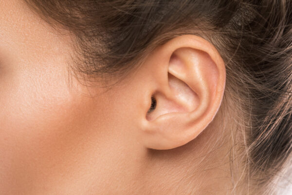
- The outer ear consists of the pinna (also called the auricle), ear canal and eardrum.
- The middle ear is a small, air-filled space containing three tiny bones. These are called the malleus, incus and stapes, but collectively they’re called the ossicles. The malleus connects to the eardrum linking it to the outer ear. The stapes (smallest bone in the body) connects to the inner ear.
- The inner ear has both hearing and balance organs. The hearing part of the inner ear is called the cochlea. This comes from the Greek word for ‘snail’ because of its distinctive coiled shape. The cochlea, which contains many thousands of sensory cells (called ‘hair cells’), is connected to the central hearing system by the hearing or auditory nerve. The cochlea is filled with special fluids which are important to the process of how the ear works.
The central hearing system consists of the auditory nerve and an incredibly complex pathway through the brain stem and onward to the auditory cortex of the brain.
Diagram of the main parts of the peripheral hearing system

Get our e-newsletter
Stay up to date with the latest news, stories and updates from Hearing Link Services and from across the deaf sector.
How do we hear?
The physiology of how our ear works, just like its anatomy, is very complex indeed. It is best understood by looking at the role played by each part of our hearing system described above.
Sound waves
These are vibrations in the air around us, are collected by the pinna on each side of our head and are funnelled into the ear canals. These sound waves make the eardrum vibrate. The eardrum is so sensitive to sound vibrations in the ear canal that it can detect even the faintest sound. It can also replicate even the most complex of sound vibration patterns.
The eardrum vibrations caused by sound waves move the chain of tiny bones (the ossicles – malleus, incus and stapes) in the middle ear transferring the sound vibrations into the cochlea of the inner ear.
This happens because the last of the three bones in this chain, the stapes, sits in a membrane-covered window in the bony wall which separates the middle ear from the cochlea of the inner ear. As the stapes vibrates, it makes the fluids in the cochlea move in a wave-like manner. This stimulates the microscopically small ‘hair cells’.
Hair cells
Remarkably, the ‘hair cells’ in the cochlea are tuned to respond to different sounds based on their pitch or frequency of sounds. High-pitched sounds will stimulate ‘hair cells’ in the lower part of the cochlea. Low-pitched sounds will stimulate ‘hair cells’ in the upper part of the cochlea.
What happens next is even more remarkable. When each ‘hair cell’ detects the pitch or frequency of sound to which it’s tuned to respond, it generates nerve impulses. These travel instantaneously along the auditory nerve.
These nerve impulses follow a complicated pathway in the brainstem before arriving at the hearing centres of the brain, the auditory cortex. This is where the streams of nerve impulses are converted into meaningful sound.
All of this happens within a tiny fraction of a second … almost instantaneously after sound waves first enter our ear canals. It is very true to say that, ultimately, we hear with our brain.
What’s happening when you have problems with your hearing?
Hearing well depends on all parts of our auditory system working normally. This is so that sound can pass through the different parts of the ear, to the brain to be processed without any distortion. The type of hearing problem you have depends on which part of your auditory system is not responding well.
Examples of potential issues
If you have a problem in the outer or middle ear, it means that there is inefficient transfer of sound to the cochlea in the inner ear. Generally, this affects the volume of sound so that it simply doesn’t seem loud enough.
A typical example would be the effect of a blockage of wax in the ear canal or a perforated eardrum. This is called a conductive hearing loss because sound vibrations are not being conducted efficiently. The cochlea is still working normally but simply not receiving enough information via its connection with the middle ear.
If the problem is somewhere between the cochlea in the inner ear and the brain, this is called a sensorineural hearing loss (SHL). The pathway through the outer and middle ears is functioning normally but, after sound arrives at the cochlea, it isn’t processed normally. This is either because of damage to the delicate ‘hair cells’ in the cochlea or to the auditory nerve. Or, because of defects in the auditory pathway leading to the brain.
There are many causes of SHL, but exposure to excessive noise or the effects of ageing are the most common. The typical signs of SHL are a general difficulty in hearing clearly and problems understanding speech in difficult listening conditions such as in background noise.
It is also possible to have both conductive and sensorineural hearing loss. This is generally called a mixed hearing loss.
For more detail about types of hearing loss, see our causes of hearing loss page.
Video of how the ear works
Courtesy of MED-EL
Webpage updated: April 2024